How do cells acquire their identities? In hopes of answering this question, a Duke team recently completed a study explaining the expression of stem cells after a decade of research.
Stem cells are found in every living organism, including humans and plants. They are initially unspecialized and can develop into almost any cell type while dividing to produce new cells over time. At this point, they’re faced with a choice: divide to make copies of themselves or create something new.
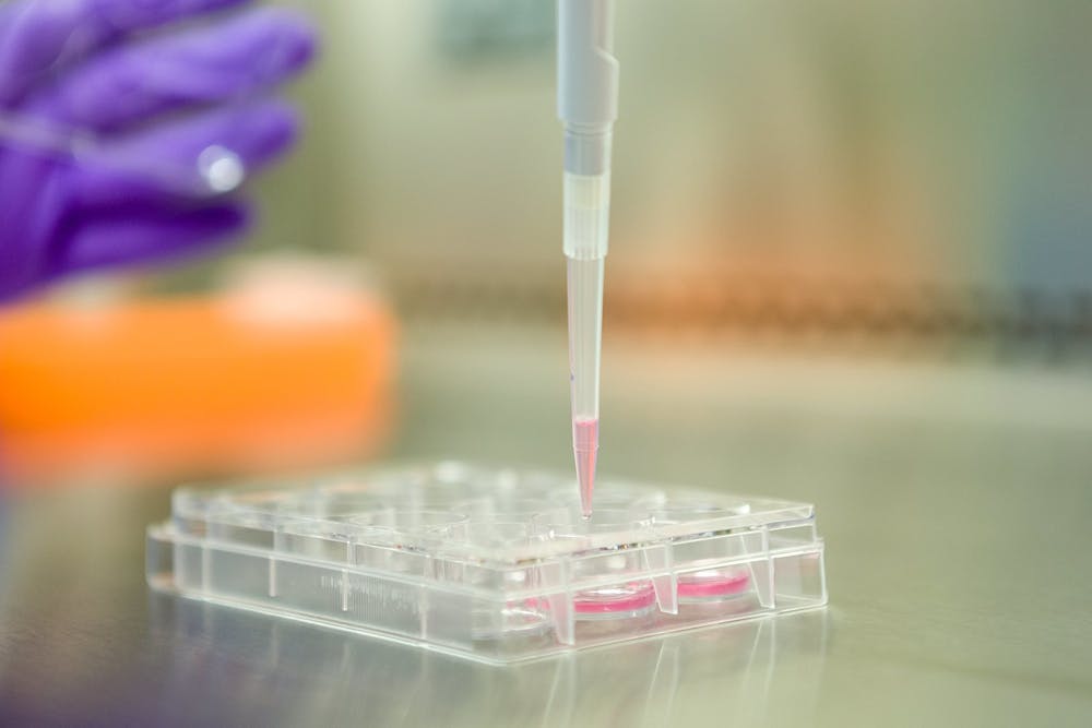
To explore how stem cells make this decision, the group researched the expression of stem cells in plants by developing a specialized microscope that takes precise photographs through a technique called light sheet microscopy.
Cara Winter, associate research professor in biology, and postdoctoral associate Pablo Szekely began working on the project in the lab of Philip Benfey, former Paul Kramer distinguished professor of biology who passed away last year.
This eventually grew to a collaboration with the California Institute of Technology, which included a microscopist, a computer scientist, graduate students and undergraduate student assistants. Winter emphasized that the project was a collaborative effort.
“There's no one person that knows all of the information that you need to know to make the microscope and the project work. So you really have to work together to figure out how to get to the goal,” Winter said.
Researchers at the California Institute of Technology visited the Duke lab to help construct the microscope. Compared to traditional imaging, this specialized light sheet microscope caused less toxicity and photobleaching, which allowed the team to image cells for longer. This was key because the microscope needed a long observation period to collect enough data on the cells.
The microscope tracked the changing colors of the root cells, which indicated whether the cell would divide and the type of division that would occur. The researchers then connected this data with the level of proteins in the cells to explore how those proteins were associated with cell division. The level of high-resolution imaging of stem cells over such a significant period has never been done before.
Szekely noted that this was made possible by using “machine learning and other technological tools to answer these biological questions.”
The study allowed the team to photograph the expression of multiple genes in the context of a living organism and connect that gene expression to cell division. They were also able to use that data to test a model of a gene regulatory network, which allowed them to gain insight into asymmetric cell division and how it interfaces with the cell cycle.
The group’s results were published in Nature. The study's findings connect to further research for humans and other animals in cancer therapies, drugs, and other aspects of the cell cycle.
“I'm hoping to continue following with the next steps of this project, following up on sort of the ideas that came out of the paper, trying to understand exactly what these two regulators are doing early in the cell cycle,” Winter said. I'm hoping to stick with imaging, continuing imaging and developing new imaging technologies to ask questions that couldn't be asked.”
Get The Chronicle straight to your inbox
Signup for our weekly newsletter. Cancel at any time.
Aseel Ibrahim is a Trinity first-year and an associate news editor for the news department.