The $10 microchip that allows your smartphone to take a panoramic shot can replace 3-D medical imaging technology worth thousands of dollars, researchers at Duke have discovered.
Existing ultrasound machines with 3-D imaging capabilities can cost up to $250,000. However, Duke researchers have found a new way to upgrade 2-D ultrasound machines at a relatively low cost. The brainchild of emergency physician Joshua Broder, the inexpensive 3-D imaging device is made possible through the use of orientation technology found in cell phones.
Broder, director of the emergency medicine residency program at the School of Medicine, explained that the microchip is attached to the ultrasound probe using a 3-D printed holster.
“We are using, in a novel way, existing chip technology. In every smartphone, there are chips that provide orientation information at a very high rate of speed. What we’ve done is piggybacked one of those chips onto an ultrasound probe,” Broder said. “The magic is that a little tiny bit of data that’s seemingly not that sophisticated all of a sudden opens up this dramatic possibility of three-dimensional imaging of the human body.”
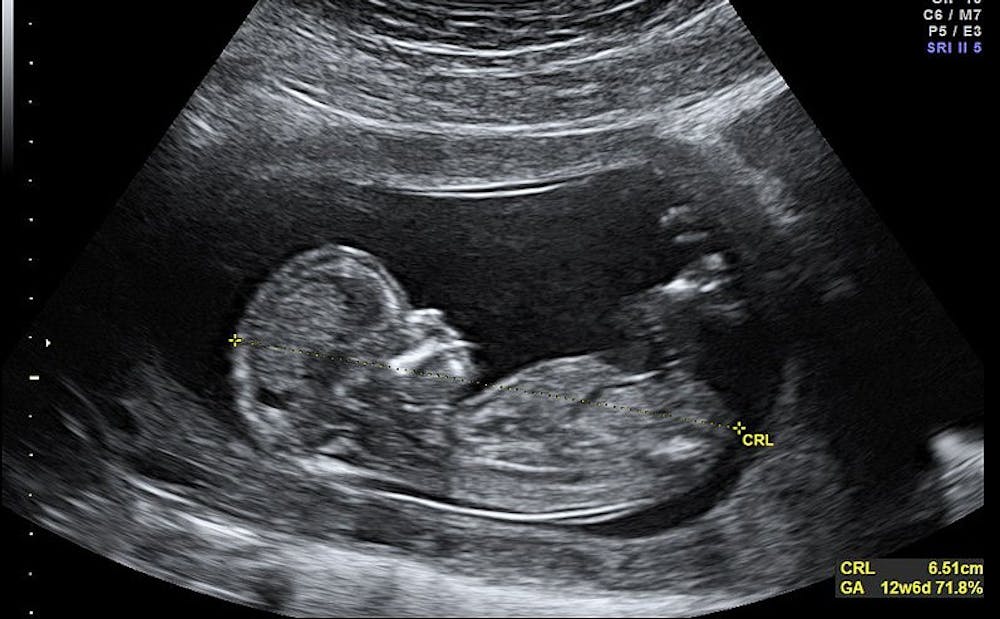
Traditional 2-D ultrasound images do not have information about the direction or angle that the probe is facing when the images are taken, Broder said. With this device, every image generated by the machine is tagged with the orientation of the probe, so that the 2-D images can be assembled into a 3-D picture.
Matt Morgan, a graduate student at Duke who worked on the device as an undergraduate, wrote in an email that the 2-D ultrasound image is a “slice” of the 3-D anatomical volume. He compared 2-D ultrasound imaging to trying to map out a living room in the dark using just a flashlight.
“You know there are couches, a coffee table, and a dining room table around somewhere, but you can only look at a small fraction of the room at any one time, so it could get confusing to figure out what the layout of the room is without any prior knowledge,” Morgan wrote. “What we’re trying to do is let you flip the lights on, so you can see the whole room simultaneously.”
Broder noted that the 3-D capability gives much needed “context” to existing 2-D images, which he explained is especially helpful in fields like emergency medicine. He added that the device will also improve patient-care by decreasing exam time.
“From a patient stand-point, if the physician can just sweep the ultrasound probe across the area that we’re interested in, in a matter of seconds, we can increase the patients’ comfort,” Broder said.
Alongside Broder and Morgan, Carl Herickhoff and Jeremy Dahl, who are former biomedical engineering instructors and professors at Duke, also helped design the device. Since then, they have both moved to positions at Stanford University where they have begun clinical trials for the device. Herickhoff said he hopes to have physicians using the device in clinics at Stanford very soon.
In addition to its low cost, another benefit of the device is its versatility, Herickhoff noted.
“We developed it to the point where it was versatile enough to plug in to any existing ultrasound system,” he said.
Since many clinics already have access to 2-D ultrasound machines, the developers predicted that the inexpensive and versatile device will be extremely popular once its reaches the market.
“By giving health care providers the ability to see the whole field of view by acquiring 3-D images instead of the conventional 2-D images, we hope to make point-of-care ultrasound more complete and intuitive at a very low cost,” Morgan wrote.
Broder, Morgan, Dahl and Herickhoff have an international patent pending on the design.
Get The Chronicle straight to your inbox
Signup for our weekly newsletter. Cancel at any time.