A team of Duke researchers recently designed a handheld probe capable of imaging the retinas of children in higher definition than ever before.
Joseph Izatt—Michael J. Fitzpatrick professor of biomedical engineering—conducted the study and incorporated new technologies from different fields of study. Francesco LaRocca, Pratt '11, who also received his Ph.D. from Duke in 2016, was the principal author of the study, which included researchers from the Pratt School of Engineering and the Duke Eye Center who helped craft the novel device. Researchers hope the high-definition imaging will be able to better identify the onset of certain vitreoretinal diseases and study juvenile retinal development.
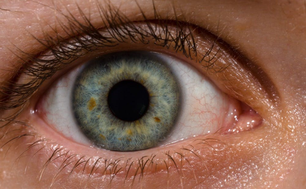
“Children have eye diseases just like adults do, which damage their retinas in various ways, and so it’s important to be able to diagnose the damage to their retina so that doctors can diagnose the disease and then try to develop a treatment for [the children],” Izatt said.
Izatt noted that in the past, imaging had been limited to mostly adults and deceased children.
Bulky machines requiring stillness and intense focus on the part of children are likely to fail because of a child’s limited attention span, LaRocca explained.
“Children have difficulty fixating, so you can’t use traditional tabletop systems," LaRocca said. "[A device] has to be small and lightweight for the clinician to get good images from the subject."
A handheld, portable probe removes the need for intense concentration and allows clinicians to image the retinas of children more effectively.
LaRocca added that providing crisp images down to the photoreceptor level will allow doctors to identify a disease in its early stages, while also permitting researchers to analyze how the density of juvenile photoreceptors changes over time.
Not only will the device be able to perceive the onset of certain retinal diseases, but it may also yield clues for developing preventative measures.
“Genetic diseases affecting photoreceptors are of particular interest because they progress to loss of visual function. The eye is a ‘relatively contained organ’ with reasonably easy access,” wrote Cynthia Toth, Joseph A.C. Wadsworth professor of ophthalmology, in an email.
Toth added that the eye is good subject for research to treat disease due to its ease of access and the ability to measure outcomes.
The handheld device was the by-product of past researchers’ efforts, combined with more recently developed technology. LaRocca explained that after first attempting to develop a small handheld device for retinal imaging, he received feedback from doctors that the machine still exceeded the ideal size.
This response led him to develop three additional modifications: a single light source instead of two, a compact micro-electromechanical systems scanner and a converging beam design. Together, these adjustments allowed the machine to be reduced to the size of a quarter.
Although the design is novel, it is unlikely that the current version of the device will become available to doctors, Izatt said.
After being used for a year at Duke Clinic, where 20 to 30 patients were imaged, the innovation has improved retinal imaging of younger patients.
"[The current version] was used mostly for getting our feet wet and having the clinicians use it and give us feedback so that we can generate the next device," LaRocca said.
LaRocca said he is optimistic that after thorough trials at Duke, the next wave of retinal imagers would catch the eye of a company specializing in the manufacturing of such devices. Then, patients would be able to benefit from the extra precision and ease of examination.
The researchers noted that they are looking forward to refining the probe and continuing to make improvements.
"One direction we've taken was to create a very large field of view," LaRocca said. "That would be very clinically useful for imaging other things, not necessarily photoreceptors, but other retinal diseases as well."
Get The Chronicle straight to your inbox
Signup for our weekly newsletter. Cancel at any time.