Duke scientists’ recent breakthrough in brain mapping technology has enabled them to make an improved model of a mouse’s brain that can be used to help determine the origins of neurological disorders and improve treatment in humans.
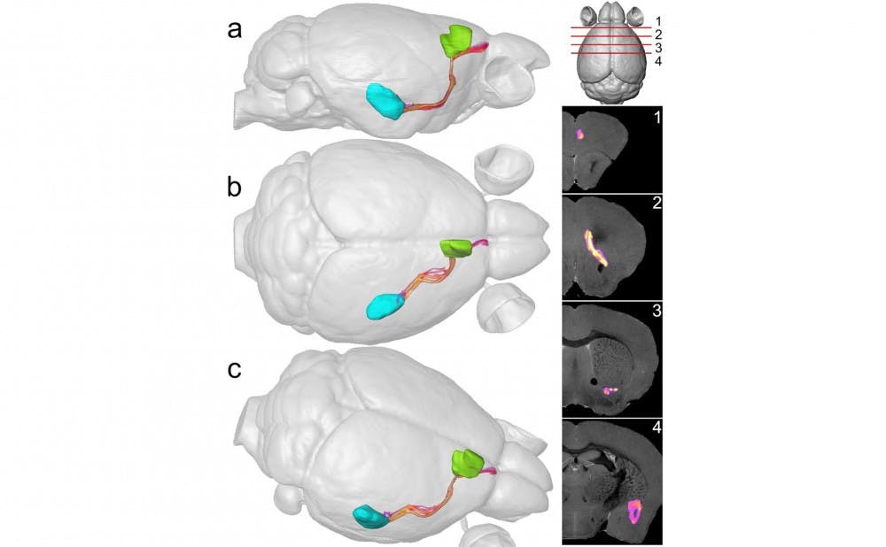
A team at the Center for In Vivo Microscopy has succeeded in creating a three-dimensional map of the circuitry inside of a mouse’s brain—known as as a connectome—at a resolution 100,000 times greater than that of a clinical magnetic resonance imaging scan. Such a rendering of the brain allowed the scientists to determine how different regions are connected and the strength of these connections at a greater precision than ever before. This in turn has far-reaching implications in the medical community, especially for targeting specific areas of the brain for treatment of diseases.
“There’s a lot of work with small animals to study diseases that’s ongoing in our lab and many other places. What’s new that we do—what we have done since 1986—is we have worked to get higher resolution than anybody else,” said senior author G. Allan Johnson, director of the Center for In Vivo Microscopy and professor of radiology, biomedical engineering and physics.
The model was created by taking the dead brain tissue of one mouse, running an advanced MRI scan on it for two weeks and then using super-computers to track the diffusion of water along the neuron wiring pathways in the mouse brain. Johnson noted that what sets their efforts to map the mouse brain apart from the others is how high the resolution is and the fact that it comes from just one mouse.
Standard microscopy procedures for mapping connectomes include cutting very thin slices of brain tissue, running them under water and physically watching the diffusion across pathways, Johnson explained. This exhaustive and invasive approach requires samples of different brain regions from almost 1,300 mice in order reconstruct a complete map. Instead, this latest technique uses just one brain from one mouse, left intact, so that the image generated is inherently three dimensional and allows for a greater ability to maneuver the model, test different pathways and compare the brains of various mice.
“Let’s take a mouse model of autism, schizophrenia, dementia—almost any neurological disease. We can probe to test where in the wiring of these mouse models there are connectivity differences,and then apply this to humans, who are much more difficult to examine,” Johnson said. “People vary so much, and they’re alive and they’re people. You can’t do a study at 100,000 times the spatial resolution in a human being, but now you have indications of where in the patient population you should be looking.”
Although researchers have already begun making an atlas of the human brain, it cannot be done with nearly the same precision as with mouse models, he noted. So far, the highest resolution of a human brain is just 1,000 times greater than that of a conventional MRI. When studying diseases, rather than struggling to interpret the lower resolution of human scans, scientists can simply examine a mouse's connectome to determine a general region as the starting point.
Connectomes at this level of precision are already helping surgeons direct the implantation of electrodes, which send impulses along various wiring connections of the brain to treat conditions such as tremors, Parkinson’s and Alzheimer’s—just to name a few.
“Still, there are limitations,” Johnson noted. “First is the ambiguity of the connectivity that we generate, because even at our high level of spatial resolution, it still isn’t high enough to resolve a single axon. Instead, we have to determine connections probabilistically, on a super computer. Second, is the amount of time it takes—two weeks to scan and another two to analyze it.”
Johnson remains optimistic, however, because this new method of mapping still significantly improves on the level of ambiguity and the length of the entire process. He is also hopeful that the engineers in his lab will be able to reduce the scan time by a factor of four or five within the next year.
“The high-resolution fiber pathways not only can serve as an atlas to delineate the intricate brain connectivity, but will also be used as a roadmap to further guide the technological development for in vivo brain imaging in translational applications,” Allen Song, director of the Center for Brain Imaging and Analysis, wrote in an email.
Get The Chronicle straight to your inbox
Signup for our weekly newsletter. Cancel at any time.